")
")


![]()


The INM (Montpellier Neuroscience Institute) imaging facility has been part of the regional imaging facility "Montpellier Ressources Imagerie" (MRI: www.mri.cnrs.fr) since its creation in 2004. It gathers scientific and technical skills dedicated to imaging in neurosciences. In addition, the facility is equipped with various cutting-edge microscopy devices for fixed or living samples, as well as image analysis solutions. It is certified ISO 9001:2015 and labeled IBISA (Infrastructures Biology Health & Agronomics: www.ibisa.net). The facility is open to external public and private laboratories.
Our roles include:
- Providing microscopy and image analysis workstations
- Assistance in the preparation and observation of samples
- Development and adaptation of new in vitro and in vivo applications
- Automation of image processing
- Training and teaching
- Expertise
1. MULTIPHOTON MICROSCOPY
Zeiss LSM 7MP / OPO
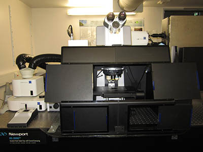
Multiphoton microscope on an upright stand Zeiss Axio Examiner. System equipped with a pulsed laser Coherent Chameleon Ultra II (wavelength range: 690 to 1010 nm) coupled to a Coherent Compact Optical Parametric (wavelength range: 1050 to 1300 nm). 4 detectors: 2 conventional PMT NDD and 2 PMT BiG with photon counting option. Environmental control chamber. ZEN software.
This microscope is dedicated to the following multiphoton imaging applications:
- Multidimensional and multicolor in vivo imaging
- Small animal imaging
- Fluorescence, Second Harmonic Generation, Third Harmonic Generation
- Coherent Anti-Stokes Raman Spectroscopy (CARS)
2. LIGHTSHEET MICROSCOPY
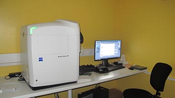
ZEISS LightSheet Z1
LightSheet microscope entirely automatized for fast 3D imaging os small living organisms (embryo like) or cleared samples. Microscope with an illumination arm and a detection arm with a 5x or 20x objective, and 2 sCMOS cameras. Illumination sources: lasr diodes 405 nm, 488 nm, 561 nm; possible Far Red detection. ZEN software.
- In vivo and cleared samples imaging
- Multidimensional and multicolor imaging
- 360° sample rotation (multiview)
- Incubation chamber
3. CONFOCAL MICROSCOPY
ZEISS LSM 880 Airyscan
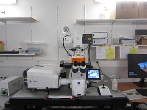
Laser scanning confocal microscope with one pinhole, on an inverted stand Zeiss Axio Observer. Detection range adjustment: several nanometers. 3 PMT detectors including one with GaAsP cathod, more sensitive. Airyscan module to increase SNR and resolution, Fast Airyscan option. Illumination with lasers or diode lasers: 405nm, 440nm, 458nm, 488nm, 514nm, 561nm, 633nm. ZEN software.
- Fixed samples
- Multidimensional and multicolor imaging
- High resolution imaging
- Fully motorized system
ZEISS LSM 700
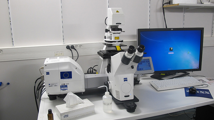
Laser scanning confocal microscope with one pinhole, on an inverted stand Zeiss Axio Observer. Adjustment of detection range: filters + secondary dichroic beamsplitter (VSD: Variable Secondary Dichroic from 400 to 700 nm). 2 PMT detectors. Illumination sources: laser diodes 405nm, 488nm, 555nm, 639nm. ZEN software.
- Fixed samples
- Multidimensional and multicolor imaging
- Fully motorized system
4. WIDE FIELD MICROSCOPY
ZEISS AXIO IMAGER Z2 - module APOTOME 2.0
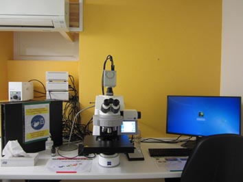
Motorized microscope on an upright stand Zeiss Axio Imager Z2, equipped with an Apotome module allowing optical sectionning in fluorescence and a sCMOS camera Hamamatsu Orca flash 4.0 LT (Black & White, 16 bits, high quantum efficiency). Available filters: Dapi/Hoechst, GFP, Cy3, dsRed, Cy5. ZEN software.
- Fixed samples between slide and coverslip
- Optical sectioning: Apotome module
ZEISS 'Cell Observer' AXIO OBSERVER
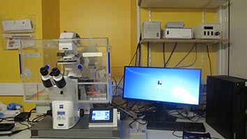
Motorized microscope on an inverted stand Zeiss Axio Observer, sCMOS camera Hamamatsu Orca flash 4.0 LT (Black & White, 16 bits, high quantum efficiency). Environmental control chamber. White light for transmission; diode Colibri for fluorescence. Available filters: Dapi/Hoechst, GFP, Cy3. Sample holders for: slides, Petri dishes, multiwell plates, … ZEN software.
- Fixed and live samples
- Time laspes (until several days)
ZEISS AXIOVERT 200M (not-MRI)
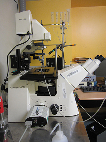
Workstation developped on an inverted stand Zeiss Axiovert 200M, a fast wavelength switch (Sutter Lambda DG-4) and a camera CoolSnap ES. The system allows to observe the response of several neurons simultaneously, with a 3-4 frames per second frequency. MetaFluor software.
- Live samples
- Calcium imaging
- Medium perfusion
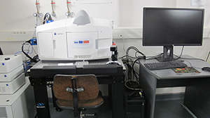
5. HIGH CONTENT SCREENING
HAMAMATSU NANOZOOMER
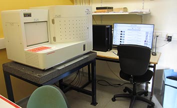
High content screening imaging system, dedicated to immunohistochemistry staining (RGB image). Automated acquisition until 210 slides. Possible fluorescence. NDP Scan software.
- Histological staining
- Service level agreement for numerization of slides (fluorescence or brightfield)
ZEISS AXIOSCAN Z1
High content screening imaging system, mainly dedicated to immunofluorescence (camera Hamamatsu Orca flash 4.0, Black & White), possible immunohistochemistry staining acquisition with RGB camera Hitachi. Automated acquisition until 100 slides. 7 LEDs available: 385nm, 435nm, 475nm, 515nm, 555nm, 590nm, 635nm. ZEN software.
- Batch of immunofluorescence slides
6. MACROSCOPY
Stereomicroscope Zeiss Discovery.V20 (no MRI) to observe big samples (as small organism, pour observation de gros échantillons (petits organismes, whole tissue sections…); available image acquisition. Wide field of view and long working distance. Axiovision software.
- Bright field
- Dark field
- Fluorescence
- LED transmitted illumination
- LED reflected brightfield illumination
7. IMAGE PROCESSING
Workstation dedicated to image processing, powerful enough to process images of several tens of GB. Available softwares: Imaris, Fiji/ImageJ, Volocity, Huygens, Definiens. The facility particularly offers its expertise for image processing and analysis with Definiens.
- Deconvolution
- Visualization and/or 3D reconstruction
- Quantification
- Segementation
- Colocalization
- Tracking