")
")


The Electron Microscopy Platform (COMET) of the Institute for Neurosciences of Montpellier (INM, Université de Montpellier/INSERM) is accessible to the different teams of INM and is also widely open to external laboratories. It has a Transmission Electron microscopy (TEM) and a Scanning Electron Microscopes (SEM).
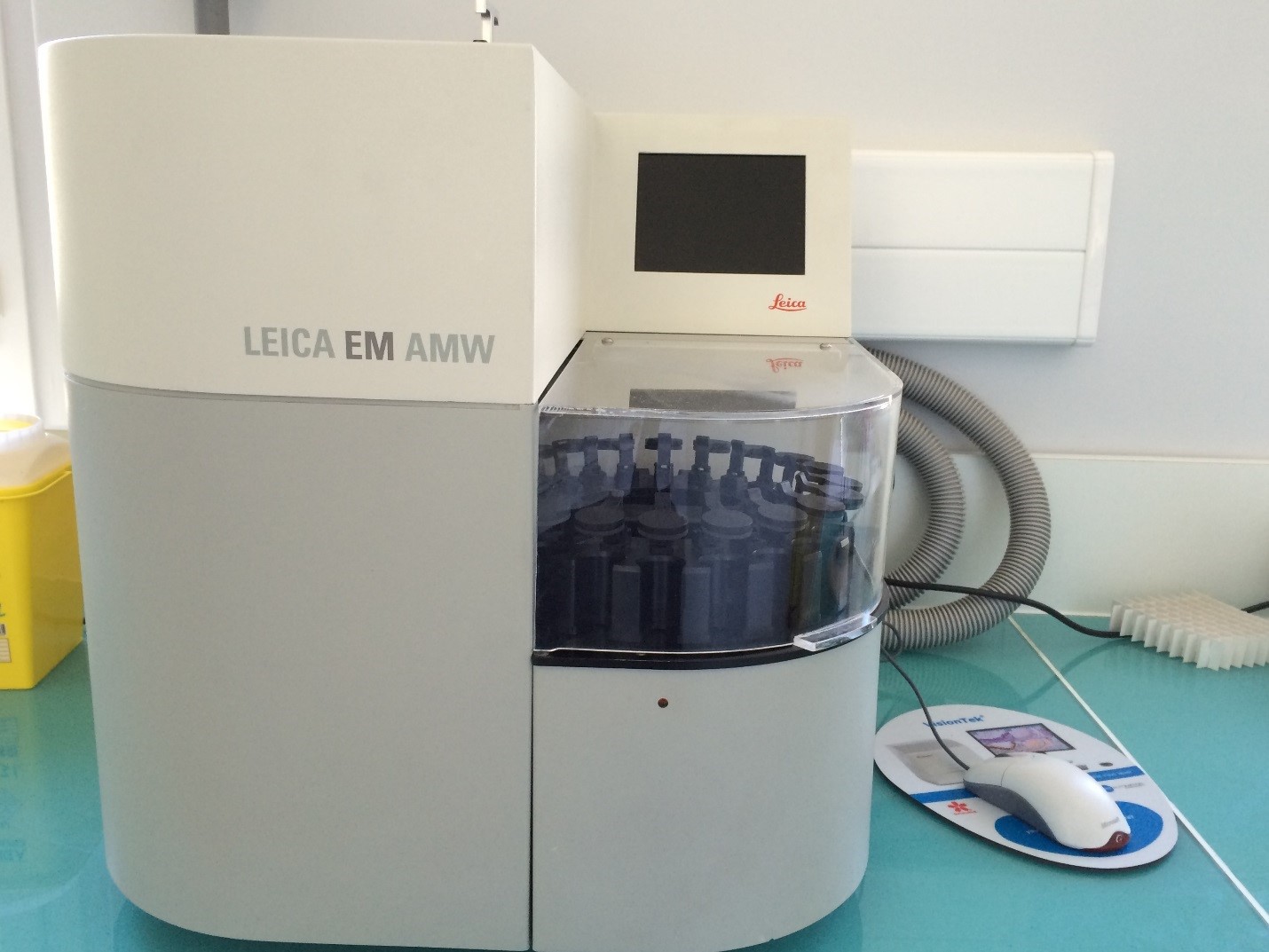
Users of the platform not only have an access to both EMs equipment, but they are assisted for preparation of their specimens: post-fixation, semi-thin and ultra-thin sections gold sputter coating; critical point, They are also tutored, if needed, during observations, analyses and interpretations.
Transmission Electron Microscopy (TEM)
Preparation:
Post-fixation (Osmium), Embedding in epoxy resin
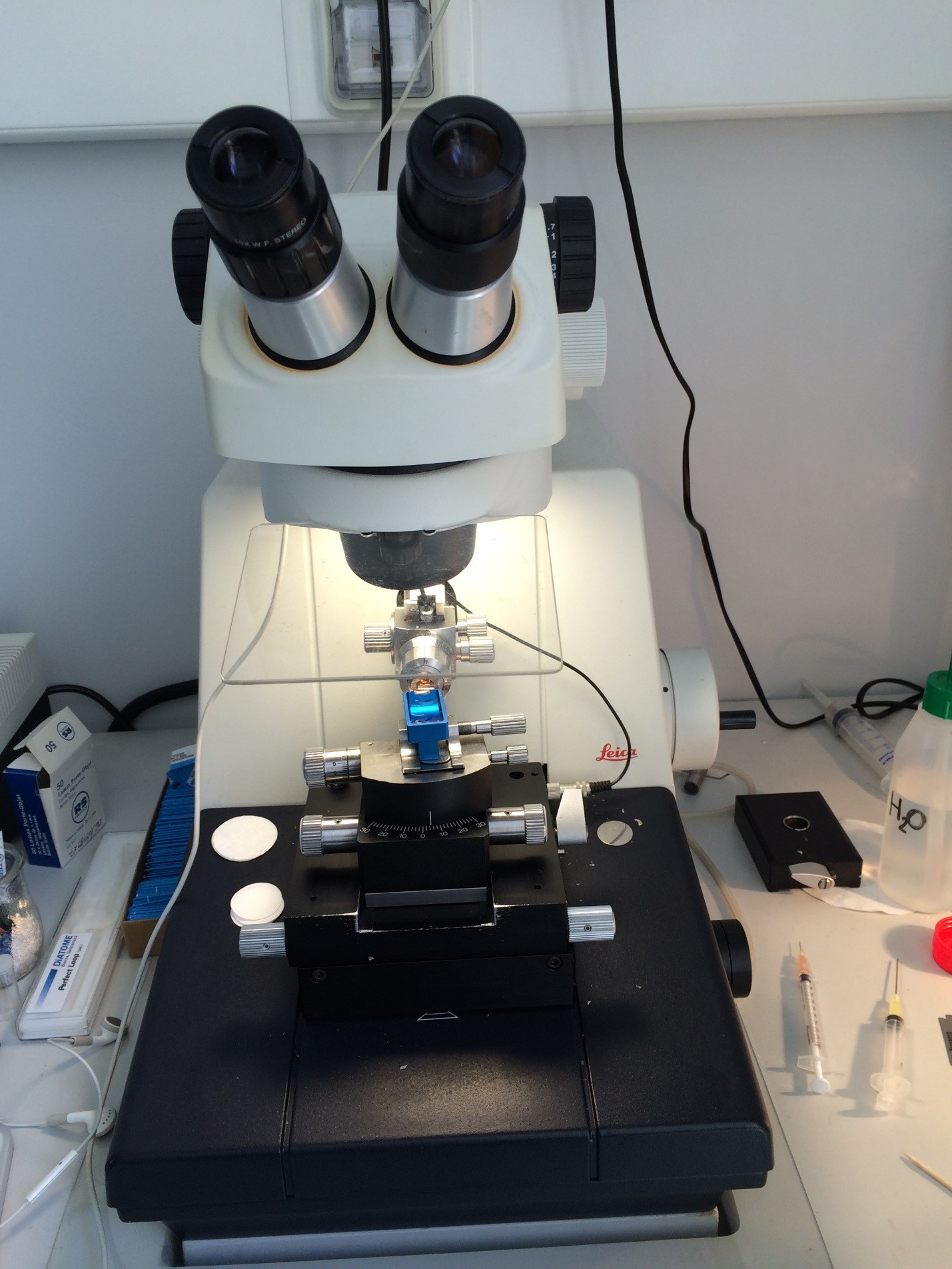
Ultramicrotomy : Semithin section (700nm) and ultrathin sections (70nm for 2D observation or 350 nm for tomography)
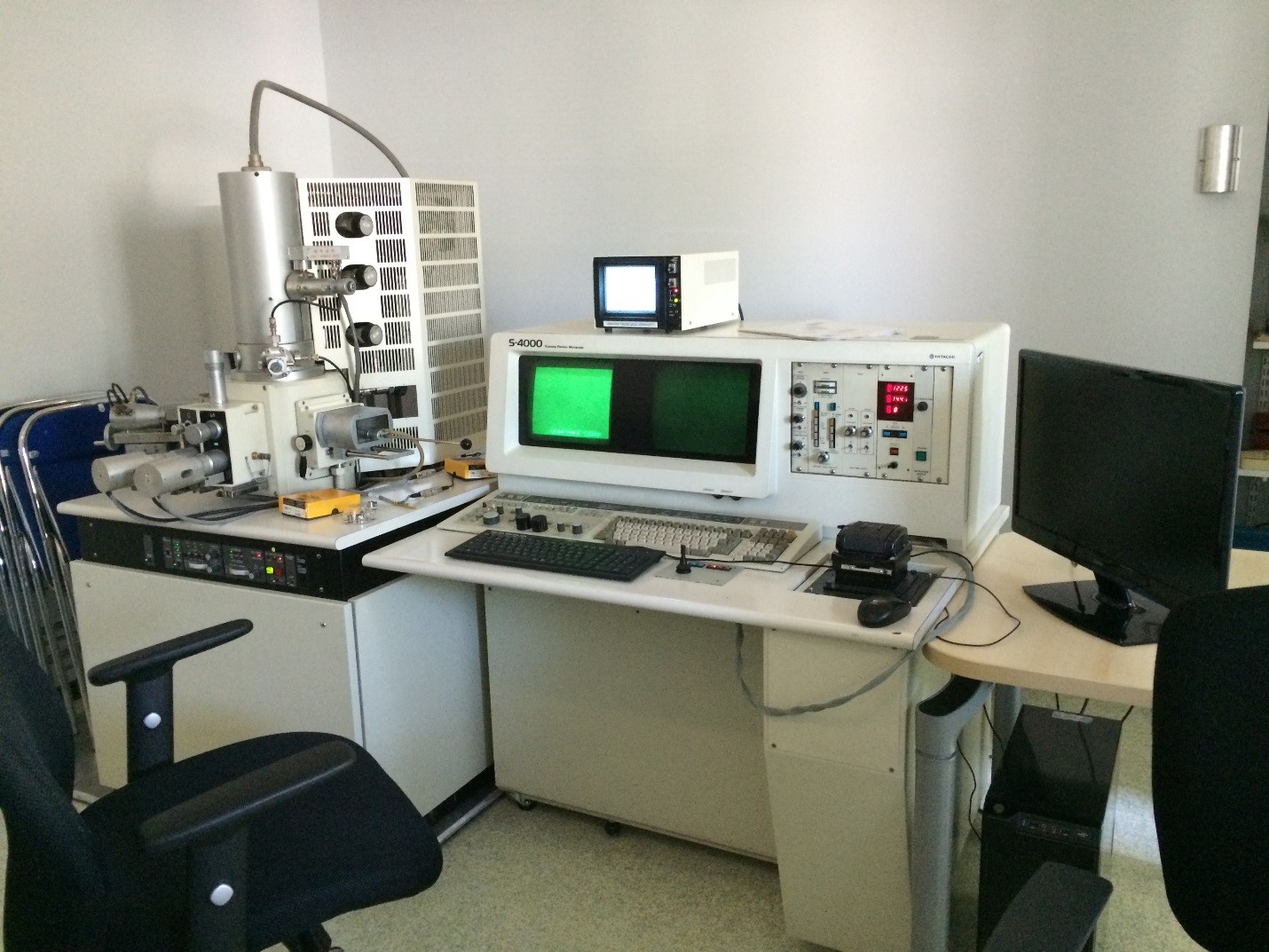
Equipment: Tecnai FEG 200KV

Classical (2D) TEM: observation of slices (70nm thick, contrasted Uranyl Acetate + Lead Citrate
TEM Tomography: slices 300-400 nm (possibility to Tilt the grid from +60° to -60°1 image every °).
In development: with serial tomography, a 3D-reconstruction can be performed on structures over 1 µm.
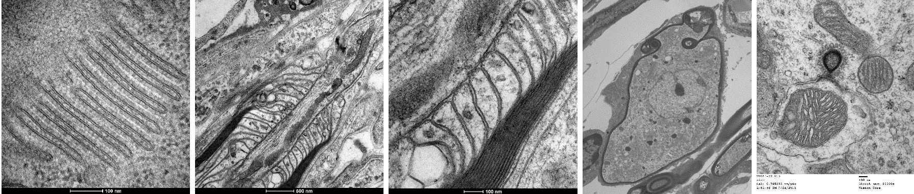
A B C D E
A: Double plasmic membrane and ribosomes. C. elegans organel. Rémy Pujol.
B and C: Myelin before a Ranvier node (mouse). Rémy Pujol/Anne-Gabrielle Harrus
D: Neuron. Guinea pig cochlear ganglion. Remy Pujol.
E: synapse ribbon and mitochondria. Mouse utricle. Remy Pujol.
Scanning electron microscopy (SEM)
Preparation:
Critical point, gold sputtering
Equipment: Hitachi S4000
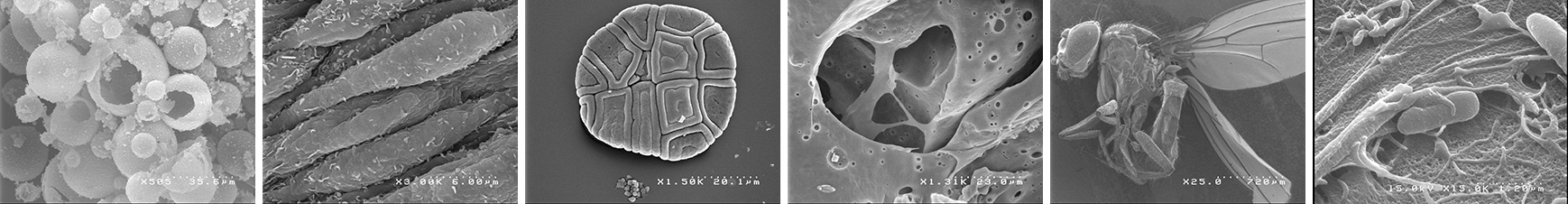
A B C D E F
A: silica granules
B: epithelial cells of the umbilical cord artery
C: mimosa pollen
D: vegetal wall
E: drosophila.
F: cultured lung cells that phagocyte Burkolderia cepacia
Contact